

Patients are often shown a low arch, a high arch, or a pronating foot and told, 'That is why you hurt.' It is a compelling story because the shape is easy to see. It is also incomplete. Foot posture influences mechanics, but research does not support treating a static arch as a verdict on pain, injury, or athletic potential.
After repeated ankle sprains, the useful question is not whether the foot looks textbook-perfect. It is whether the rearfoot and midfoot adapt under load, the intrinsic and extrinsic muscles control that motion, the ankle has enough dorsiflexion, and the whole system can sense the ground and produce force during the task that exposes the problem.
The short answer
Foot posture is one examination finding, not a diagnosis or a treatment plan.
Pronated, neutral, and high-arched feet can all be pain-free, strong, and athletic. Static posture does not reveal whether the foot is adaptable, sensitive, coordinated, or able to create force. Research links posture with selected movement and injury variables, but the effects are too small and condition-specific to label one shape defective.
In chronic ankle instability, posture can influence available motion or balance. That makes it worth testing; it does not justify forcing every arch higher or putting every patient in the same orthotic. The intervention has to change the patient's actual movement, symptom, or performance test.
Separating structure from performance
Static alignment does not determine how the foot will perform under load.
The arch changes with body size, load, joint mobility, bone shape, ligament properties, muscle activity, age, and the position used for measurement. A static Foot Posture Index describes alignment; it does not directly measure how the foot responds during running, landing, or fatigue.
Systematic review evidence suggests pronated posture is associated with a small increase in risk for some overuse conditions, not all lower-extremity injury. Newer work also shows that static posture does not necessarily correlate with dynamic balance. Those findings argue for context rather than correction by appearance alone.
Shape
What the arch and rearfoot look like in a chosen position.
Mobility
How the foot changes between non-weight-bearing and loaded positions.
Function
How it senses, adapts, produces force, and supports the task.
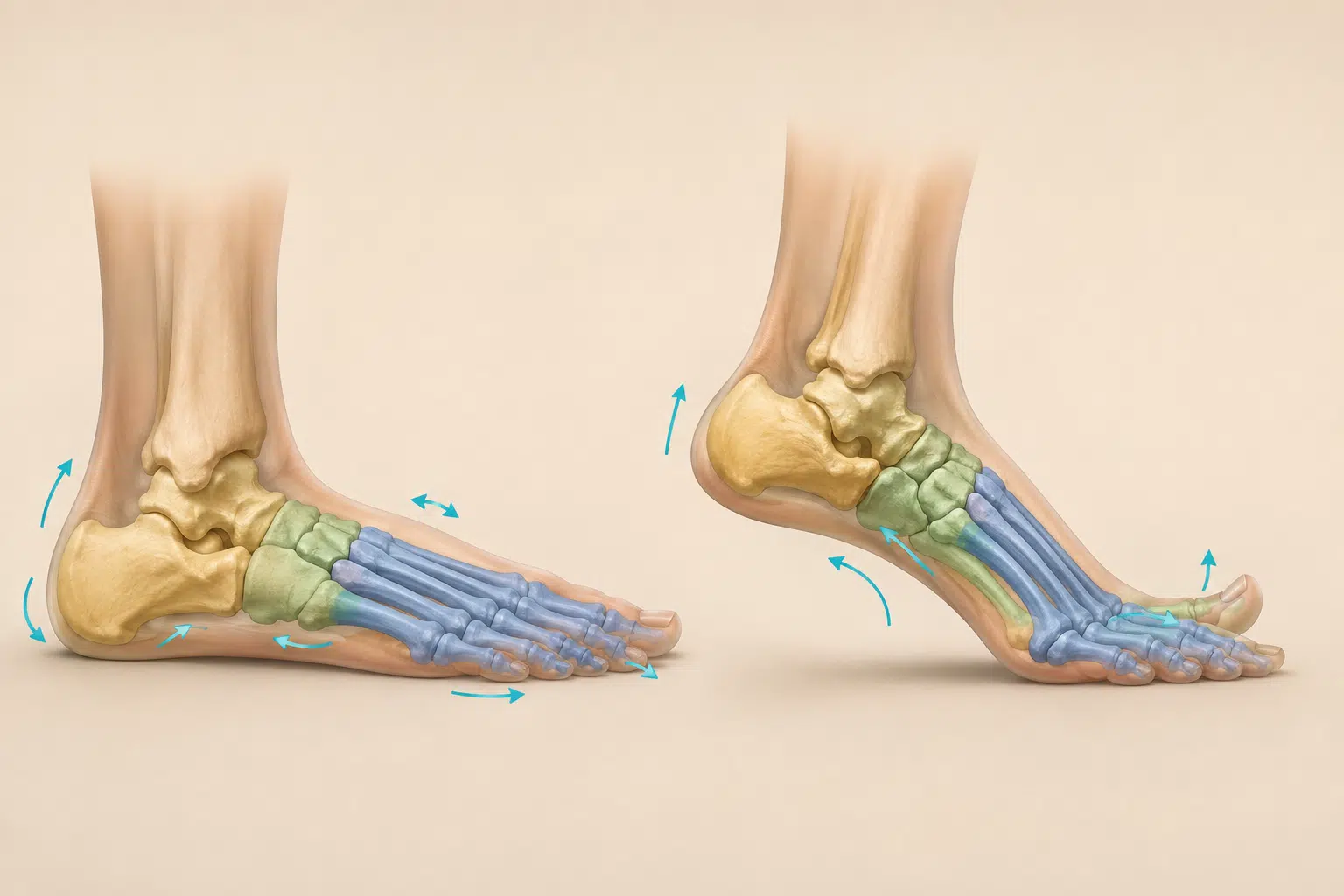
How the foot and ankle share motion
Rearfoot, midfoot, forefoot, and ankle motion combine during weight-bearing tasks.
The rearfoot centers on the talus and calcaneus and includes talocrural and subtalar motion. The midfoot includes the talonavicular and calcaneocuboid region plus the navicular, cuboid, and cuneiform joints. The forefoot includes the metatarsals and toes, with the metatarsophalangeal joints becoming especially important as the heel rises.
During loading, these regions move in three planes and do not follow one identical rotation. Rearfoot eversion, tibial advancement and rotation, midfoot deformation, forefoot spreading, and arch lowering can occur with different timing and magnitude. During later stance, pressure advances toward the forefoot, the heel rises, the toes dorsiflex, and plantar tissues plus active muscle help increase foot stiffness for propulsion.
Pronation and supination are useful composite descriptions, not single-joint diagnoses. Midfoot motion is not automatically compensation, and the foot does not become a completely rigid block at push-off. The clinical question is whether the person's segmental strategy is comfortable, controllable, and adequate for the force and speed of the task.
Anatomy in Motion uses pronation as a 'mobile adaptor' and supination as a 'rigid lever' to teach this shift from adaptation toward propulsion. That is a useful clinical teaching model, especially because it treats pronation as necessary rather than defective. It should not be read as proof that every bone follows one fixed sequence in every person; direct multi-segment studies show meaningful motion throughout the foot and substantial individual variation.
- Heel position. Does the heel stay grounded and move in a way the person can control?
- Midfoot mobility. How much does the arch change, and is that change symptom-related?
- Forefoot and toes. Can the big toe and forefoot accept load and support propulsion?
- Ankle dorsiflexion. Is the talocrural motion itself limited when foot compensation is controlled?

Foot function after repeated ankle sprains
The foot contributes to ankle stability alongside calf strength, position sense, balance, and proximal control.
People with chronic ankle instability can show differences in balance, proprioception, dorsiflexion, gait, and rearfoot alignment. Those group findings do not prove the same deficit exists in every individual. They identify what deserves measurement.
A foot that cannot sense or manage load reduces the quality of the base beneath the ankle. But calf strength, peroneal and tibial muscle capacity, hip strategy, visual input, surface, footwear, fatigue, and prior exposure all contribute to stability. A short-foot exercise is not a complete ankle program.
- 01
Measure the foot in load.
Compare posture, mobility, symptoms, and control rather than relying on appearance.
- 02
Test the ankle separately.
Dorsiflexion, strength, tenderness, swelling, and instability deserve their own measures.
- 03
Challenge the system.
Single-leg reach, heel raise, hop, landing, and sport tasks reveal different demands.
- 04
Change one variable.
Foot cue, footwear, support, mobility, or exercise is re-tested against the original complaint.
A task-specific foot and ankle examination
Structure, motion, force, balance, footwear, and the relevant task should be interpreted together.
The clinician compares Foot Posture Index, navicular or midfoot mobility, big-toe motion, weight-bearing dorsiflexion, heel-raise quality and endurance, intrinsic foot control, balance, gait, footwear, and the specific athletic or daily task. None of these tests is a universal score of good or bad feet.
An intervention earns emphasis when it changes a meaningful sign. If a foot cue improves balance or a squat but has no effect on running symptoms, the next test should be closer to running. If an orthotic or shoe helps, it can be a useful option without implying the foot is structurally broken.
When a focal calf or lower-leg symptom blocks ankle motion, dry needling can be tested against the same weight-bearing movement. A positive response lowers the symptom barrier; it does not change foot shape or replace the strength, balance, and reactive work that rebuilds stability.
Training adaptation and force production
Foot exercises should progress from isolated control to loaded balance, propulsion, landing, and sport.
Systematic reviews and trials support intrinsic foot muscle training for selected measures of foot function, balance, and patient-reported outcomes. The evidence is heterogeneous, and visible arch remodeling is not required for meaningful improvement.
Training can begin with finding a tripod contact and controlling the arch without toe gripping. It must progress to heel raises, single-leg loading, reaching, gait, jumping, and the surfaces or footwear the person actually uses. The ankle, calf, hip, and larger kinetic chain are trained alongside the foot.
Sense
Improve awareness of pressure under the heel, first metatarsal, and fifth metatarsal.
Control
Coordinate arch, toes, rearfoot, ankle, and calf without rigid gripping.
Load
Use the foot during heel raises, balance, landing, running, and sport-specific work.
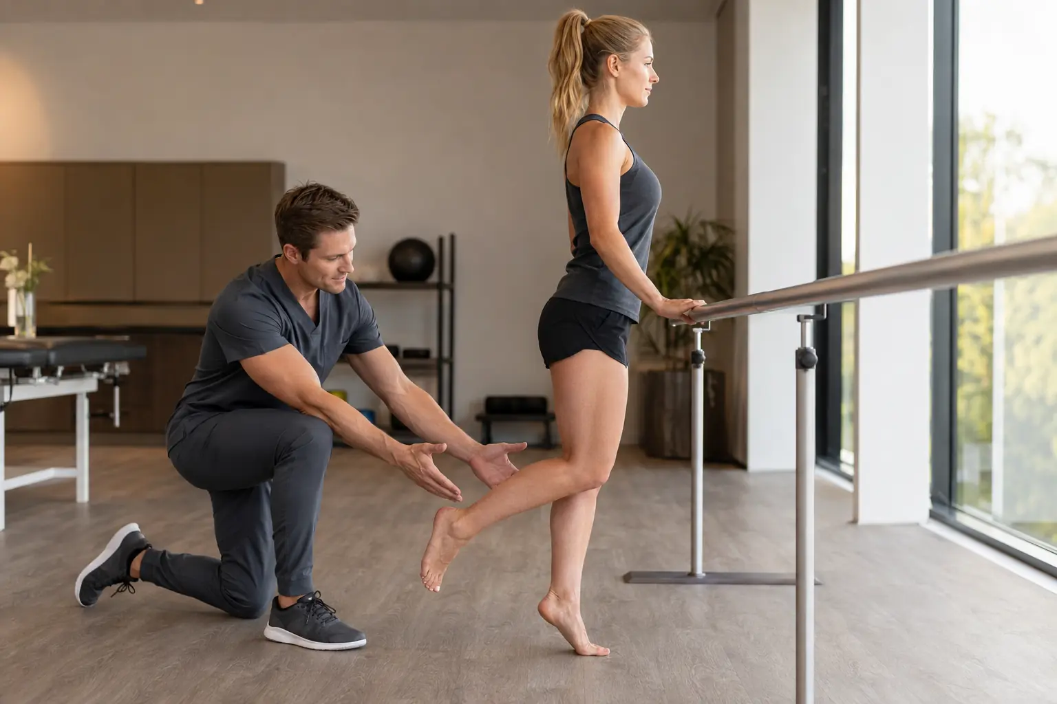
What confident care looks like
Foot structure does not need to look ideal for mobility, balance, and strength to improve.
A useful plan respects anatomy without turning normal variation into damage. It tests the foot as a dynamic part of the lower extremity and trains the qualities connected to the person's actual limitation.
When the right variable changes balance, mobility, or symptoms, that response gives the program direction. The goal is a foot-and-ankle system that can adapt to the ground and create force with confidence.
Continue exploring
Choose the next question that fits your symptoms.
Why the ankle still feels stiff after a sprain
Connect foot function with dorsiflexion, calf capacity, and chronic instability.
Read nextWhy an old injury still feels tight
Understand how healed tissue and incomplete function can coexist.
Read nextMovement assessment
See how findings are tested against symptoms and real activity.
Read nextCommon questions
Practical answers for the next step.
Are flat feet bad for ankle stability?
Not automatically. Many people with low arches are strong and symptom-free. Foot posture can influence mechanics or risk in specific contexts, but static appearance alone does not determine instability or pain.
Can high arches contribute to repeated sprains?
Certain foot alignments can influence loading and balance, but repeated sprains are multifactorial. A clinician should also assess dorsiflexion, strength, proprioception, balance, footwear, fatigue, and task exposure.
Can foot exercises change my arch?
They can change muscle function, control, balance, and selected arch measures. A visible structural transformation is not required for improved symptoms or performance.
Do I need orthotics?
Not everyone does. Orthotics can be a useful symptom or load-management option for selected people. Their value should be judged by comfort, function, and the task response rather than the idea that every foot must be corrected.
Is short-foot exercise enough for chronic ankle instability?
No. It can be one component, but comprehensive rehabilitation usually includes ankle mobility, calf and surrounding muscle strength, balance, reaction, hopping or landing, and progressive return to activity.

Written & clinically reviewed by
Dr. Brandon Lim, PT, DPT, CMTPT, FMSC
Dr. Lim is the founder of Limitless Physiotherapy in Columbia, Maryland. His one-on-one approach integrates detailed examination, movement assessment, manual therapy, dry needling, and progressive exercise according to each person's presentation and goals.
Meet Dr. LimSelected sources
Evidence behind the discussion.
- Hogan KK, et al. Influence of foot posture on dorsiflexion and postural control in chronic ankle instability. Clin Biomech. 2016.
- Neal BS, et al. Foot posture as a risk factor for lower-limb overuse injury: systematic review and meta-analysis. J Foot Ankle Res. 2014.
- Jaffri AH, et al. Intrinsic foot muscle training for foot function and dynamic postural balance: systematic review. J Athl Train. 2023.
- Lee DR, Choi YE. Six-week intrinsic foot muscle exercise in chronic ankle instability: randomized trial. J Exerc Rehabil. 2019.
- Origo D, et al. Foot Posture Index does not correlate with dynamic postural stability. Sports. 2024.
- Leardini A, et al. Rear-foot, mid-foot and fore-foot motion during the stance phase of gait. Gait Posture. 2007.
- Lundgren P, et al. Invasive in vivo measurement of rear-, mid- and forefoot motion during walking. Gait Posture. 2008.
- Kelly LA, et al. Active regulation of longitudinal arch compression and recoil during walking and running. J R Soc Interface. 2015.
- Ward G. Part 2—The Foot: Anatomy in Motion educational model of triplanar pronation and supination. Podiatry Review. 2022.