

Leg symptoms can be unsettling even when the lower back barely hurts. A long drive produces burning through the buttock and calf. Standing at a counter brings on heaviness or tingling. Bending to tie a shoe sends an electric sensation toward the foot.
Those differences help decide what to test first. The examination compares strength, sensation, reflexes, symptom behavior, and the way walking, sitting, bending, or changing position affects the familiar leg symptom. That is far more useful than choosing a stretch from a symptom map.
The short answer
“Sciatica” is a starting word—not a complete diagnosis.
The sciatic nerve forms from several lower-spine nerve roots and travels through the pelvis and leg. In everyday conversation, almost any pain reaching the buttock or leg may be called sciatica, but research has used the same word for several different conditions.
A 2023 working group from the International Association for the Study of Pain recommended the broader term spine-related leg pain, with further description of whether the presentation is somatic referred pain, radicular pain, or radicular pain with radiculopathy. Patients do not need to memorize those terms. The distinction matters because pain alone, nerve-related pain, and objective loss of nerve function do not carry exactly the same priorities.
Three related patterns
Where the pain travels is only one part of the picture.
These patterns can overlap. Pain quality and location may offer clues, but no drawing, dermatome chart, or single test can classify every person correctly.
Somatic referred pain
Lower-back, pelvic, or hip structures can produce buttock or leg pain without a spinal nerve root being the primary source. When no neurologic involvement is present, strength, reflexes, and sensation are usually intact.
Radicular pain
Abnormal activity in a spinal nerve root or related structures may feel electric, shooting, burning, sharp, or tingling. Disc herniation, inflammation, or narrowing can contribute, but a pain map cannot prove the level or cause.
Radiculopathy
Objective loss of nerve-root function can include reduced sensation, measurable weakness, or a changed reflex. It can occur with radicular pain, but the terms are not interchangeable.
Pain location starts the examination; strength, sensation, reflexes, symptom behavior, and movement response tell us which pattern fits.
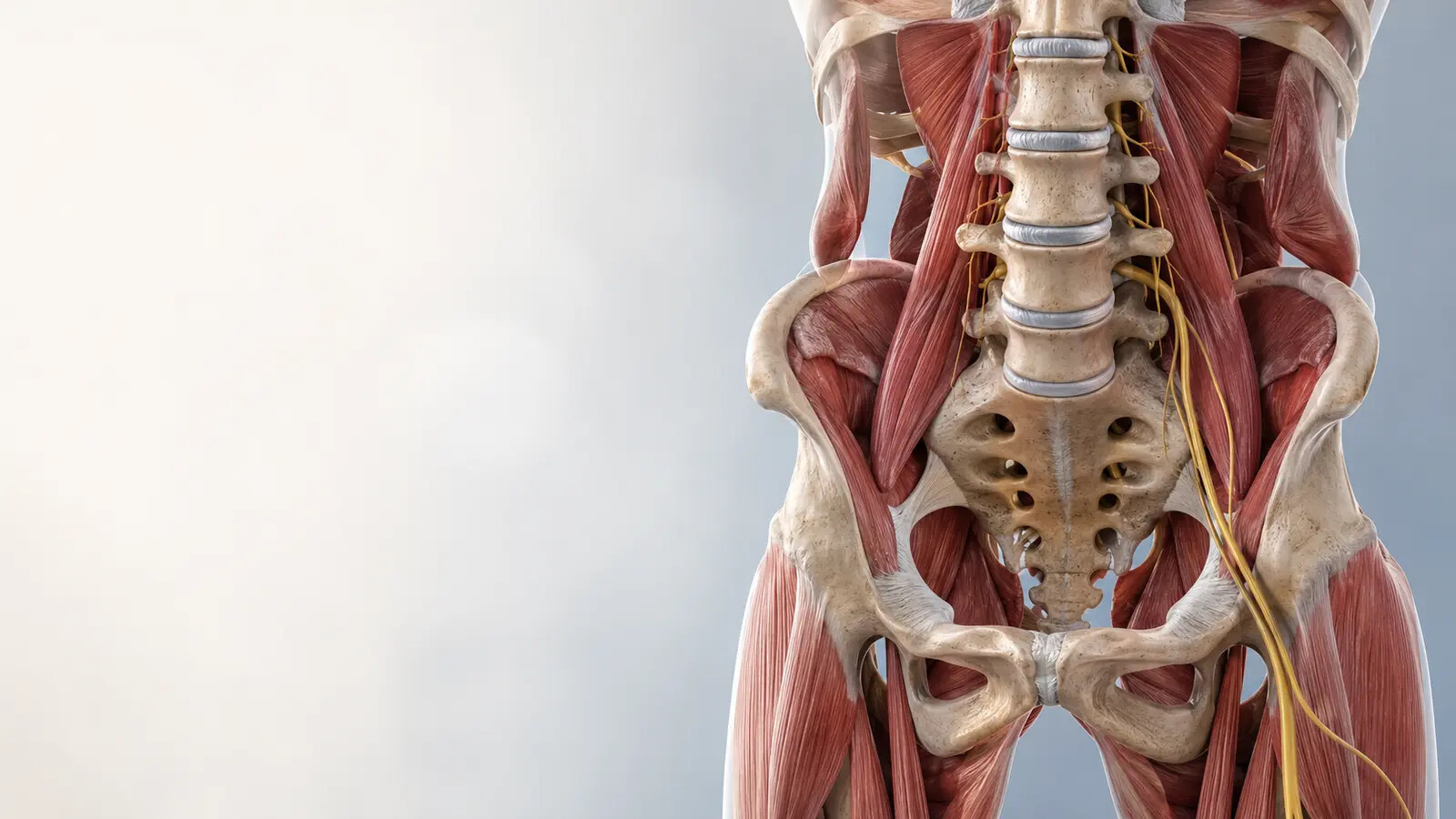
Possible sources and mimics
Several conditions can create a similar path into the leg.
A lumbar disc and nearby nerve root
A disc herniation can irritate or compress a lumbar nerve root. Symptoms may change with sitting, bending, lifting, coughing, sneezing, or repeated spinal movement.
Lumbar or foraminal stenosis
Narrowing around spinal nerves can produce pain, heaviness, numbness, or weakness with standing and walking. Relief with sitting or bending forward can fit this pattern but is not exclusive to it.
Referred pain from the spine, pelvis, or hip
The lower back can refer pain without radiculopathy, and hip or peri-hip conditions can overlap. Motion, loading, and symptom behavior help determine which findings matter.
Deep-gluteal or peripheral-nerve irritation
The sciatic nerve can be irritated outside the spine, and other peripheral nerves can affect the thigh, leg, or foot. A tender piriformis or relief after massage does not prove nerve entrapment.
Neither the word bulge on an MRI nor pain during one movement establishes the diagnosis. The imaging level and side, onset, neurologic findings, symptom behavior, and full examination should agree closely enough to influence a decision.
Learn more about our approach to back pain and sciatica physical therapy. When hip motion or loading appears relevant, the examination can also draw from the connected approach described in hip pain and hypermobility care.
The examination
What a useful back and leg evaluation should clarify.
No isolated maneuver is accurate enough to carry the diagnosis. A straight-leg-raise or slump test may help assess neural mechanosensitivity, but it must reproduce the relevant symptom and be interpreted with the history, neurologic findings, movement response, and other tests.
- 01
Define the onset and symptom behavior.
Trauma, distribution, and the response to sitting, standing, walking, bending, lifting, sleep, coughing, or sneezing help organize the possibilities. Work, sport, recent loading changes, medical history, and prior episodes add context.
- 02
Measure neurologic function.
Sensation, reflexes, and relevant muscle strength help identify objective nerve-root loss. Heel or toe walking, repeated calf raises, and ankle or toe testing may reveal a deficit that a general strength check misses.
- 03
Test neural mechanosensitivity without turning it into a diagnosis.
A useful neurodynamic result requires more than hamstring tightness: it should reproduce the familiar symptom and change when the nervous system is sensitized or unloaded. Even then, it is one finding in a cluster.
- 04
Observe what spinal positions and repeated movements change.
Symptoms may move farther down the leg with one direction and become more central with another. That response can guide an initial movement strategy; it does not prove that a disc has “gone back in.”
- 05
Compare the spine with the hip, peripheral nerve, and real task.
Hip motion and loading, gait, balance, vascular features, local muscular findings, and a scaled version of the meaningful task help identify a modifiable starting point.

The role of imaging
An MRI should answer a clinical question—not replace the examination.
The American College of Radiology advises that uncomplicated acute low-back pain with or without radiculopathy generally does not require immediate imaging. MRI becomes more useful when a serious condition is suspected, neurologic loss is severe or progressive, symptoms are not improving after an appropriate period of conservative care, or the result would guide an injection, surgical consultation, or another medical decision.
Imaging findings are real, but their meaning is not automatic. A systematic review of 3,110 people without pain found that lumbar disc degeneration, bulges, and protrusions became increasingly common with age. That does not make a concordant disc herniation irrelevant in someone with new leg pain and neurologic loss. It means the report must be interpreted alongside the onset, side, strength, reflexes, sensation, symptom behavior, and function.
- X-rays can help answer selected questions about bone, alignment, arthritis, or fracture, but they do not directly show a lumbar nerve root.
- MRI shows discs, nerve roots, and other soft tissues when that information is likely to change management.
- Electrodiagnostic testing may be considered when the neurologic diagnosis or level remains unclear or a peripheral-nerve condition must be distinguished from radiculopathy.
A composite clinical pattern
The same line of leg pain can require a different next step.
This example combines patterns seen in practice; it does not describe one identifiable patient. Individual presentations and results vary.
Consider an active adult whose buttock and lateral-calf pain began after a week that combined longer-than-usual driving with a sudden increase in lifting volume. Sitting and bending reproduce the familiar leg symptom. Sensation, reflexes, ankle strength, toe strength, and walking are intact. A neurodynamic test reproduces the familiar symptom and changes when neural tension is reduced. One direction of repeated movement brings the symptom out of the calf and closer to the hip, and a modified lifting setup remains tolerable the next day.
That pattern may support education, temporary modification of sitting and lifting dose, a direction-specific movement strategy, appropriately dosed neural mobility, and progressive return to loading without immediate MRI.
Now consider another person with a similar pain path who is increasingly catching the toes while walking. Objective ankle and toe strength have declined, and the weakness is worsening. The pain map may look similar, but progressive motor loss makes timely medical assessment and possible imaging more important. The line drawn down the leg did not make the decision. The neurologic change did.
Rehabilitation
Build movement options, nerve tolerance, and the capacity life requires.
There is no single best exercise for every person labeled with sciatica. A 2025 network meta-analysis of 40 randomized trials found that evidence comparing nonsurgical treatments for acute and subacute sciatica was generally of very low confidence. That uncertainty is a reason to measure the individual response—not a reason to do nothing.
In a trial of 220 adults with sciatica lasting less than 90 days, education plus four weeks of early physical therapy produced modestly greater average improvement in disability than education and usual care alone at six months. One protocol should not be treated as universal, but the result supports active, examination-based care with realistic expectations.
- Keep moving, but adjust the dose. Walking distance, sitting duration, range, load, speed, repetition, and recovery can be modified while symptoms are irritable.
- Use a directional strategy only when the response supports it. Repeated flexion, extension, side-gliding, or another movement may help one person and aggravate another.
- Restore neural mobility without aggressively stretching an irritable nerve. Neural sliders may be easier to tolerate than a sustained end-range stretch; dosage and response still matter.
- Rebuild trunk, hip, leg, and task-specific capacity. Desk work, running, lifting, and physical work require different ranges, loads, repetitions, and recovery.
- Track neurologic function and return to the real activity. Pain, distribution, walking tolerance, sleep, strength, sensation, reflexes, and next-day recovery all help judge progress.
Movement assessment and sports rehabilitation can make this progression more specific when the goal involves lifting, running, repeated bending, endurance, speed, or whole-body force transfer.
Hands-on care and dry needling
Short-term symptom change can support rehabilitation—but it does not define the diagnosis.
Manual therapy may help selected people move more comfortably, especially when it is paired with active treatment. Its value is judged by whether a meaningful symptom, movement, or task changes—not by a claim that a joint or disc was permanently put back into place.
Dry needling may be considered when a reproducible muscular component in the lower back, hip, or leg contributes to pain, guarding, or restricted movement. Reviews in chronic low-back pain suggest possible short-term pain benefit, particularly when needling is combined with other care, but those findings should not be generalized to every cause of radiating leg pain.
Dry needling does not decompress a lumbar nerve root, reverse neurologic weakness, or repair a disc. If it creates a useful short-term reduction for walking, movement, or exercise, the plan still has to build the capacity and confidence required outside the clinic.
Let symptom behavior—and neurologic function—set the pace
How much discomfort is acceptable during rehabilitation?
Some discomfort does not automatically indicate injury. A stable, tolerable symptom that settles after activity without spreading farther down the leg, increasing numbness, reducing strength, or worsening function the next day may allow gradual progression.
A progressively more distal symptom, increasing numbness, declining force, new foot slap or tripping, or a meaningful loss of walking or daily function warrants reassessment. Pain intensity matters, but a changing neurologic deficit matters more.

Safety first
When back or leg symptoms need emergency or prompt medical attention.
Seek emergency evaluation
New urinary retention or loss of normal bladder sensation, new loss of bowel control, numbness in the saddle region, or rapidly progressing weakness—especially when symptoms affect both legs—can be features of cauda equina syndrome, a rare but time-sensitive condition.
Sudden shortness of breath, chest pain, faintness, or coughing blood—particularly with a newly swollen, warm, red, or painful leg—also requires emergency evaluation for a possible blood clot.
Seek prompt medical assessment
Progressive foot or leg weakness, new foot drop, major trauma, fever or systemic illness with severe back pain, substantial fracture risk, a history that raises concern for infection or cancer, severe unrelenting symptoms, or a hot and markedly swollen leg should not be managed as routine sciatica without further evaluation.
These findings do not confirm a serious diagnosis. They change the safest next step.
Common questions
Sciatica, disc findings, and returning to activity.
Does pain below the knee mean I have a herniated disc?
No. Pain below the knee can raise suspicion for nerve-related involvement, but location alone cannot confirm a disc herniation. The history, neurologic findings, neurodynamic tests, and imaging when indicated determine how well a disc-related explanation fits.
Can sciatica occur without back pain?
Yes. Symptoms may be strongest in the buttock, calf, foot, or toes even when back pain is minor or absent. The examination should still compare lumbar nerve-root, peripheral-nerve, hip, deep-gluteal, vascular, and other possible sources.
Does a positive straight-leg-raise test prove sciatica?
No. It is more meaningful when it reproduces the familiar leg symptom and changes with sensitizing or unloading maneuvers. Hamstring tightness alone is insufficient, and the result must be combined with the history and neurologic examination.
Is piriformis syndrome the usual cause of buttock and leg pain?
No. Deep-gluteal sciatic-nerve irritation is possible, but the label is often applied too broadly. A tender piriformis does not establish nerve entrapment; lumbar, hip, peripheral-nerve, vascular, and other medical sources should also be considered.
Should I stretch my hamstring or piriformis?
Not automatically. An irritable neural structure can feel like a tight hamstring, and sustained end-range stretching may increase a spreading leg symptom. Stretching is useful only when the target, dose, and response fit the examination.
Do I need an MRI before starting physical therapy?
Often, no. Immediate imaging is usually unnecessary without severe or progressive neurologic loss, major trauma, or another red flag. MRI matters more when it could change management, recovery is off course, or an injection or surgical decision is being considered.
Can dry needling help sciatica?
It may help when a reproducible muscular component contributes to pain or restricted movement, but it does not decompress a nerve root or treat every cause of radiating leg pain. When used, it should support reassessment, exercise, and return to function.
Should I stop walking, lifting, or exercising?
Not necessarily. Temporary modification is often more useful than complete shutdown. Range, load, duration, and frequency should reflect neurologic status and the response during and after activity. Progressive weakness, numbness, or a spreading and longer-lasting leg response requires reassessment.
Does sciatica always require surgery?
No. Many people begin with nonsurgical care. Surgical consultation becomes more important with suspected cauda equina syndrome, severe or progressive weakness, or persistent disabling symptoms with a matching structural finding. Earlier surgery may provide faster relief for selected disc-related cases, while longer-term differences can narrow.
How long does recovery take?
There is no universal timeline. Some acute episodes improve over several weeks; persistent pain, stenosis, neurologic loss, recurrent symptoms, or high physical demands may take longer. Symptom distribution, neurologic function, activity tolerance, recovery, and return to specific tasks are more useful markers than a fixed deadline.

Written & clinically reviewed by
Dr. Brandon Lim, PT, DPT, CMTPT, FMSC
Dr. Lim is the founder of Limitless Physiotherapy in Columbia, Maryland. His one-on-one approach combines a detailed back, hip, and neurologic examination with movement assessment, targeted hands-on care, dry needling when indicated, progressive strengthening, and return-to-activity testing built around each person's work, training, and goals.
Meet Dr. LimSelected sources
Evidence behind the discussion.
- Schmid AB, Tampin B, Baron R, et al. Recommendations for Terminology and the Identification of Neuropathic Pain in People With Spine-Related Leg Pain. Pain, 2023.
- George SZ, Fritz JM, Silfies SP, et al. Interventions for the Management of Acute and Chronic Low Back Pain: Revision 2021. JOSPT, 2021.
- Hutchins TA, Peckham M, Shah LM, et al. ACR Appropriateness Criteria: Low Back Pain—2021 Update. Journal of the American College of Radiology, 2021.
- Fritz JM, Lane E, McFadden M, et al. Physical Therapy Referral From Primary Care for Acute Back Pain With Sciatica: A Randomized Controlled Trial. Annals of Internal Medicine, 2021.
- Zhu Z, Schouten T, Strijkers R, et al. Effectiveness of Nonsurgical Interventions for Patients With Acute and Subacute Sciatica: A Systematic Review With Network Meta-analysis. JOSPT, 2025.
- Brinjikji W, Luetmer PH, Comstock B, et al. Systematic Literature Review of Imaging Features of Spinal Degeneration in Asymptomatic Populations. AJNR, 2015.
- van der Windt DAWM, Simons E, Riphagen II, et al. Physical Examination for Lumbar Radiculopathy Due to Disc Herniation in Patients With Low-Back Pain. Cochrane Database of Systematic Reviews, 2010.
- Lin LH, Lin TY, Chang KV, et al. Neural Mobilization for Reducing Pain and Disability in Patients With Lumbar Radiculopathy: A Systematic Review and Meta-analysis. Life, 2023.
- Lara-Palomo IC, Gil-Martínez E, López-Fernández MD, et al. Efficacy of Dry Needling for Chronic Low Back Pain: A Systematic Review and Meta-analysis of Randomized Controlled Trials. Alternative Therapies in Health and Medicine, 2023.
- Peul WC, van den Hout WB, Brand R, et al. Prolonged Conservative Care Versus Early Surgery in Patients With Sciatica Caused by Lumbar Disc Herniation: Two-Year Results of a Randomized Trial. BMJ, 2008.
- American Association of Neurological Surgeons. Cauda Equina Syndrome.