
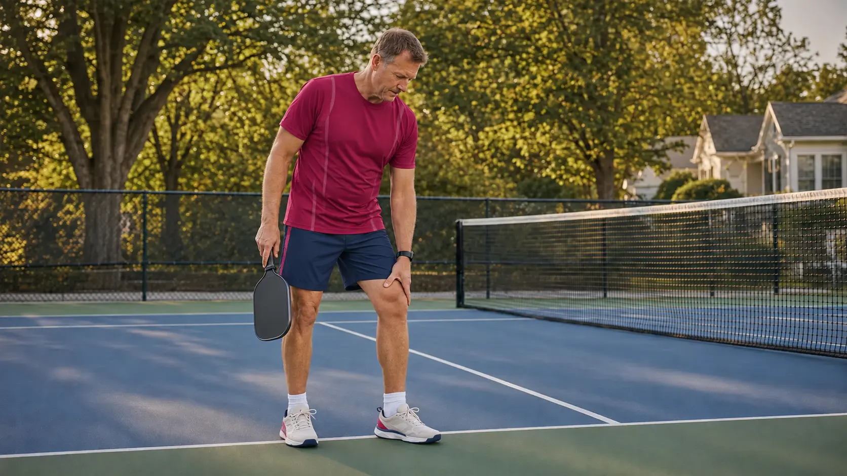
Perhaps your knee feels fine for the first few games, then begins to ache after repeated dinks and low reaches. Maybe a sharp pivot produced medial joint-line pain. Or the knee becomes stiff and puffy later that evening, even though there was no single dramatic injury.
These are not all the same problem. In a 2025 nationwide study of 1,758 U.S. players, the knee was the most commonly reported body region for a physical complaint. In a separate 2026 healthcare-system study of players who sought orthopedic care, osteoarthritis flares and meniscal tears were the two most common specific diagnoses. Those studies describe different populations and cannot tell us what is causing one person's pain—but they explain why the knee deserves a careful, sport-specific examination.
The short answer
In many non-emergency cases, pickleball knee pain reflects a mismatch between current capacity and the demands of play.
The irritated tissue may be in the knee itself: the patellofemoral joint, a tendon, the meniscus, a ligament, or an arthritic joint. The timing and amount of play may have exceeded what that tissue was prepared to handle. Hip, ankle, foot, trunk, or spine findings may also influence how force is shared—but none should be blamed from appearance alone. The useful test is whether a targeted change alters the familiar symptom or improves the court task that matters. The goal is to identify what the knee is asking for now, then build it toward repeated lunging, braking, and recovery.
Start where it hurts
Pain location helps organize the examination; it cannot settle the diagnosis by itself.
Pain location, swelling, onset, and behavior help organize the evaluation. They do not identify one structure with certainty because several knee conditions can overlap, and symptoms from the hip, back, or nervous system can occasionally be felt around the knee.
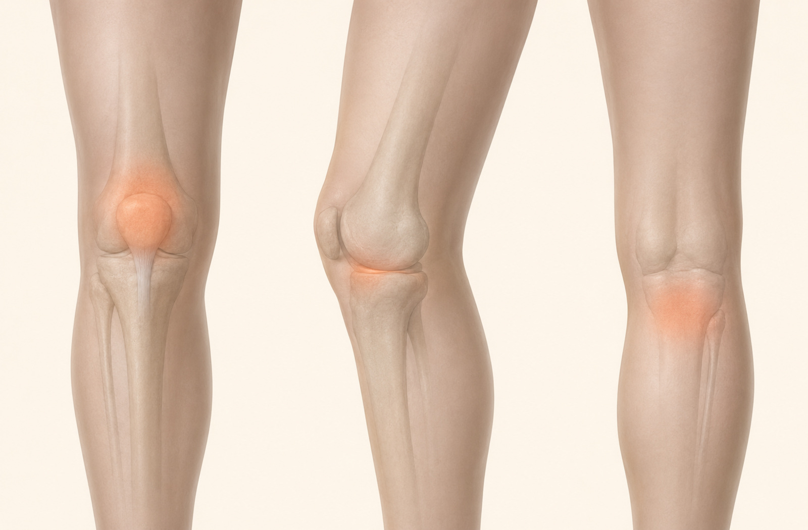
Front of the knee
Patellofemoral pain often becomes noticeable with repeated knee bending, stairs, squats, or low court positions. The patellar or quadriceps tendon may be more focal and load-sensitive.
Medial joint line
A meniscal, arthritic, MCL, or nearby tendon-related presentation may be considered. A pivot, swelling, catching, and the ability to fully bend and straighten help separate the possibilities.
Lateral knee
Patellofemoral, meniscal, ligament, tendon, or referred symptoms can overlap here. The exact movement and loading pattern matter more than the side of the knee alone.
Back of the knee
Joint swelling, a popliteal cyst, hamstring or calf tissue, and other causes can create posterior discomfort. Compare bending, straightening, calf loading, and the response after play.
The MRI deserves context
A meniscal finding must be interpreted with symptoms, swelling, motion, strength, and court demands.
A sudden twist followed by rapid swelling raises concern for a broader traumatic injury inside the knee—not only a meniscus problem—and warrants timely evaluation. Joint-line pain, loss of motion, or true mechanical locking also raises a different concern than an ache that gradually appears after longer sessions. True locking means a physical block, often preventing full extension, rather than stiffness or pain-limited motion. Clicking by itself does not confirm a meniscal tear. Meniscal findings are common on MRI in middle-aged and older adults who report no pain, aching, or stiffness.
That does not make imaging meaningless. It means the scan should be interpreted alongside the history, examination, and response to loading. For many adults with a degenerative, non-obstructive meniscal tear, exercise-based physical therapy can produce outcomes comparable with arthroscopic partial meniscectomy over the long term. Acute trauma, mechanical obstruction, significant instability, or persistent swelling may warrant a different medical pathway.
What the court asks of the knee
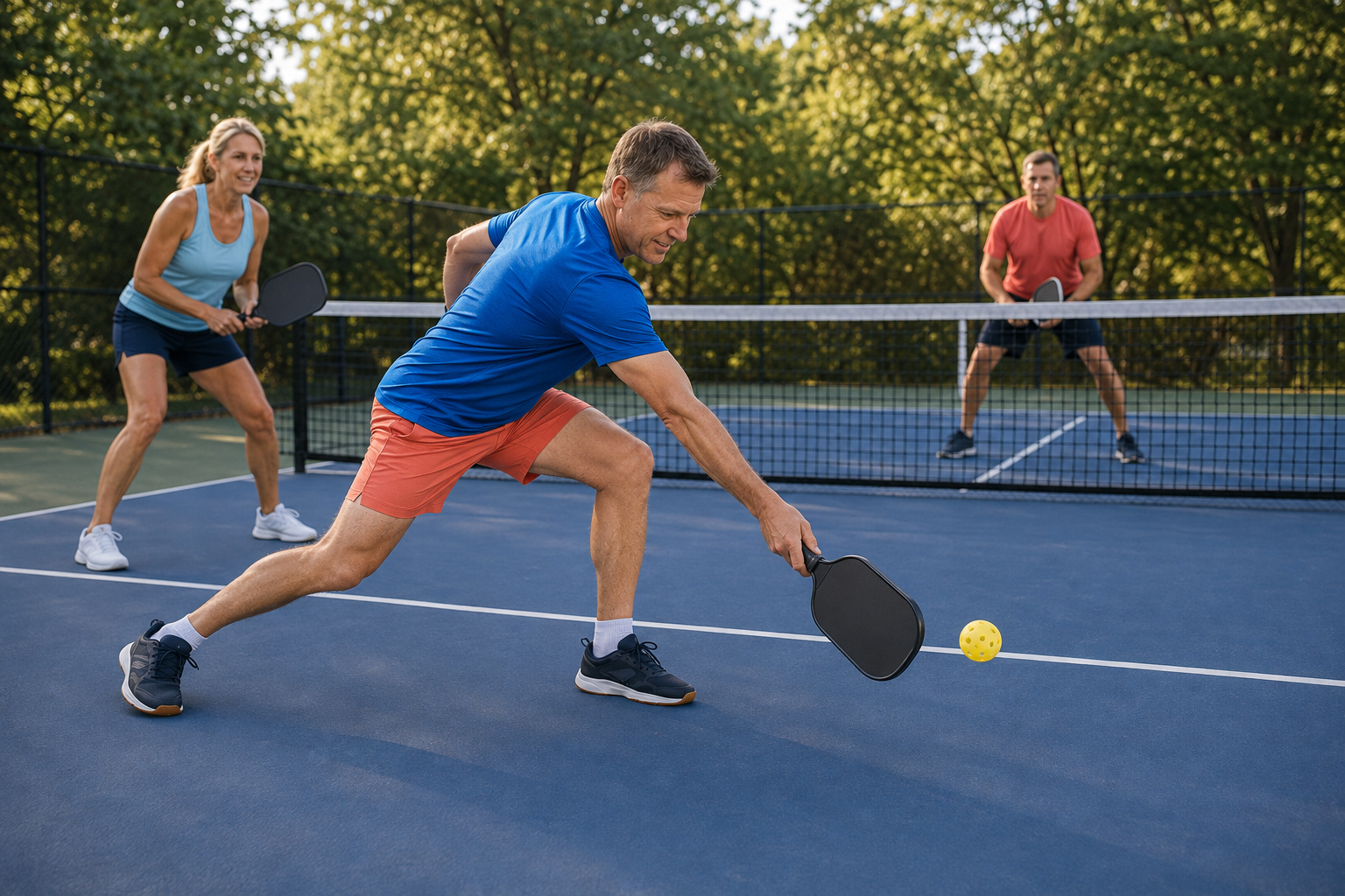
The difficult part is often not the lunge—it is controlling and leaving it.
A kitchen reach requires more than knee flexion. You have to recognize the shot, move the center of mass, place the foot, absorb forward or diagonal momentum, control the paddle, and recover before the opponent's next ball. Fatigue, a late first step, a larger-than-usual reach, or a sudden increase in playing frequency can change that demand substantially.
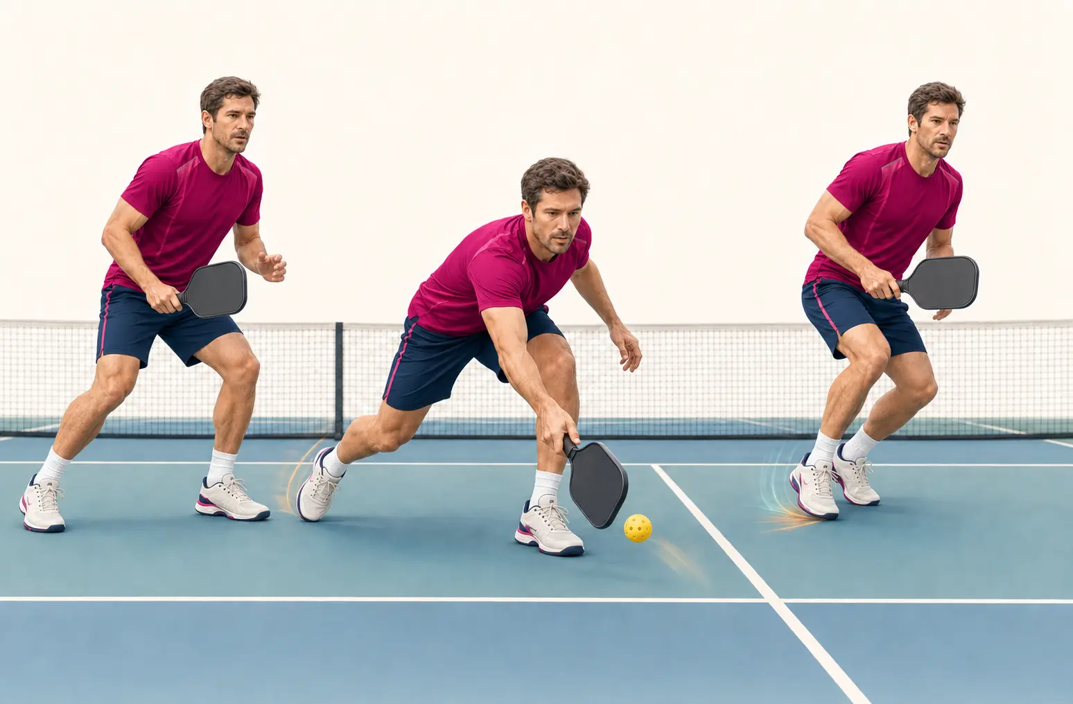
This distinction matters because common screening shortcuts can be misleading. In a study of recreational pickleball players, lunging and moving backward were the leading reported situations for falls, and players with a fall history were slower on a pickleball-specific change-of-direction test. Hip-abduction strength was not significantly different between players who did and did not report a fall, while single-leg squat form and ankle dorsiflexion were not related to fall history. The lesson is not that hips or ankles never matter. It is that a static or generic clinic test should not be treated as a diagnosis of a dynamic, reactive court problem.
This was a small cross-sectional study of 92 recreational players and relied on participants recalling previous falls. Its findings are useful clues for assessment and training—not definitive predictors of who will fall in the future.
Clinical reasoning in action
What a useful pickleball movement analysis should do.
- 01
Examine the knee first.
Onset, pain location, swelling, range of motion, joint-line and patellofemoral findings, tendon response, ligament stability, strength, and relevant neurologic or referred symptoms establish the local picture.
- 02
Reproduce the court demand.
A forward kitchen lunge, diagonal reach, lateral push, pivot, braking step, and recovery may reveal different limits. The task is observed at a tolerable speed before fatigue or reaction is added.
- 03
Test the rest of the chain without assigning blame.
Ankle motion, calf and quadriceps capacity, hip rotation and strength, foot strategy, trunk position, balance, and power are considered only when they plausibly relate to the familiar task.
- 04
Test one meaningful change against the court task.
Step length, foot placement, trunk strategy, lunge depth, external support, or a targeted mobility or strength intervention may be changed. If the familiar symptom or movement improves, that response helps prioritize the plan.
- 05
Restore speed, reaction, and repeatability.
A pain-free slow lunge is an initial benchmark. Later progression should tolerate repeated reaches, direction changes, recovery steps, decision-making, and the playing volume you intend to resume.
A composite clinical pattern
When “meniscus pain” needs a wider—and more specific—look.
This example combines patterns seen in practice; it does not describe one identifiable patient. Individual presentations and results vary.
Consider an active adult who recently increased from one casual session to three competitive sessions per week. Medial knee pain and mild swelling appear after deep kitchen reaches. An MRI report mentions degenerative meniscal change, but there was no acute pop, the knee is not locked, and daily walking is comfortable.
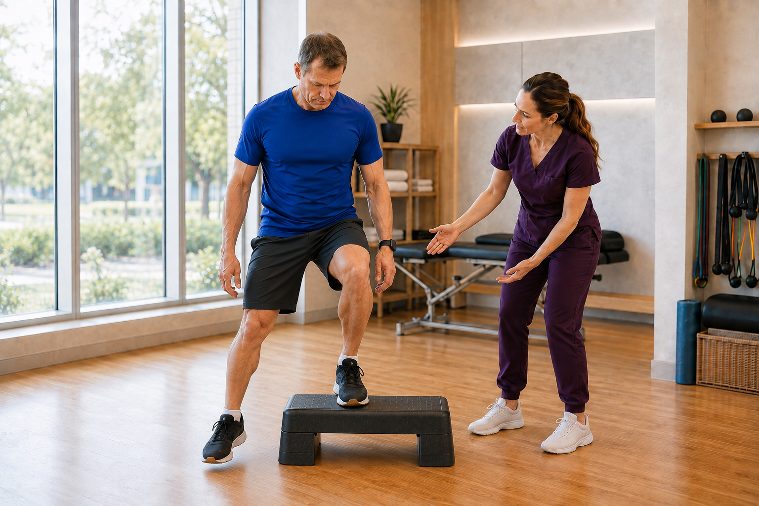
The local examination still matters: joint motion, effusion, joint-line response, strength, and stability are checked first. During a diagonal lunge, the familiar pain appears as the player arrives late, reaches from a narrow base, and struggles to brake. A small change in preparatory footwork and step length reduces the symptom during the re-test. That does not prove the movement caused the meniscal finding. It identifies a modifiable way to keep training while knee and whole-leg capacity are progressed.
Another player with the same MRI wording may have rapid swelling, a blocked range of motion, or true mechanical locking after a pivot. The scan may look similar; the appropriate next step may not be.
Build a knee that can use the court again
Rehabilitation should match the diagnosis, then exceed the demands of a generic exercise sheet.
Patellofemoral pain, patellar tendinopathy, an osteoarthritis flare, a ligament sprain, and a meniscal presentation do not all receive the same dosage or progression. Across common non-emergency knee conditions, education and appropriately dosed exercise are central. The exact plan may include temporarily modifying playing volume, restoring comfortable motion, and progressing quadriceps, calf, hamstring, hip, and trunk capacity before adding faster braking and multi-directional work.
Manual therapy can help a selected person reduce short-term pain or recover useful motion. Dry needling may be considered when a reproducible muscle-related finding in the quadriceps, calf, hamstring, or hip is limiting the next step. Neither technique repairs an arthritic joint or meniscal tear. A useful response should make it easier to move, load, and progress—not create dependence on passive care.
- Settle the flare without unnecessary shutdown. Adjust the dose of play and the most provocative movements while maintaining tolerable activity.
- Build force capacity. Progress the knee and the rest of the leg for squatting, stepping, pushing, landing, and absorbing momentum.
- Train braking and recovery. Add forward, diagonal, lateral, and backward movement in a controlled sequence before making it reactive.
- Match real playing volume. Prepare for repeated games, changing opponents, fatigue, and the speed at which technique becomes harder to maintain.
For a closer look at this approach, see movement assessment and corrective exercise and sports physical therapy and return-to-activity rehabilitation.
Use the next-day response
Can you keep playing while the knee improves?
A practical decision is based less on whether you feel anything and more on how the knee responds during play, afterward, and the next day. Stable, mild symptoms without swelling, limping, loss of motion, or declining function may allow continued modified play. Increasing pain, recurrent swelling, guarded movement, or a meaningful next-day loss of function is a reason to reduce the dose and clarify the problem.
Before play, use a progressive warm-up that rehearses the actual sport: easy forward and backward movement, lateral steps, controlled lunges, split-steps, and gradually faster direction changes. During recovery, rebuild not only slow strength but also the speed and repetition your usual games require. There is no universal “perfect” lunge and no five-exercise program that fits every painful knee.
When pickleball knee pain needs prompt medical attention.
Seek prompt assessment after substantial trauma with rapid swelling, inability to bear weight, obvious deformity, repeated giving way, or a knee that is physically blocked—especially if it cannot fully straighten. Fever, a hot and markedly swollen joint, severe unrelenting pain, or new calf swelling, warmth, and redness also require medical evaluation. Shortness of breath or chest pain with calf symptoms requires emergency care.
Common questions
Pickleball knee pain, movement analysis, and return to play.
Why does my knee hurt after pickleball but not during everyday walking?
Walking does not require the same repeated low reaches, rapid stops, pivots, lateral pushes, and recovery steps as pickleball. A knee may tolerate daily activity yet become symptomatic when the speed, depth, repetition, or total playing volume exceeds its current capacity. The pattern still needs to be examined because arthritis, meniscal symptoms, patellofemoral pain, tendon irritation, and referred pain can overlap.
Does clicking or catching mean I tore my meniscus?
Not necessarily. Clicking is common and can occur without a clinically important meniscal tear. Meniscal findings are also common on MRI in middle-aged and older adults who have no knee symptoms. A recent pivot injury, joint-line pain, swelling, or loss of motion changes the level of concern. True mechanical locking means the knee is physically blocked—often preventing full extension—rather than simply feeling stiff, painful, or hesitant to move.
Is pickleball bad for an arthritic knee?
Pickleball is not automatically harmful for a knee with osteoarthritis. Symptoms often reflect the relationship between current joint tolerance and the amount, intensity, and novelty of play. Many people can continue or return with a temporary adjustment in volume, progressive strength and conditioning, and a plan for lunging and changing direction. A large or persistent flare should still be evaluated.
Should I stop playing pickleball until my knee pain is completely gone?
Complete rest is not always necessary, but repeatedly provoking increasing pain, swelling, limping, or next-day loss of function is not a useful training strategy. Some players can continue with shorter sessions, more recovery, reduced lunge depth, or lower-intensity drilling while capacity is rebuilt. The right modification depends on the suspected condition and how the knee responds over the next 24 hours.
What should physical therapy for pickleball knee pain include?
A useful plan should examine the knee itself, screen the hip, ankle, spine, and neurologic system when relevant, reproduce the court movement that matters, and re-test after a targeted change. Rehabilitation usually progresses strength, mobility where needed, braking, lateral movement, power, and court-specific tolerance rather than stopping at a generic exercise sheet.
When does pickleball knee pain need prompt medical evaluation?
Prompt evaluation is appropriate after a significant injury with rapid swelling, inability to bear weight, obvious deformity, repeated giving way, or a knee that is physically blocked—especially if it cannot fully straighten. New calf swelling, warmth, or redness; fever; or severe unrelenting pain also requires medical evaluation. Shortness of breath or chest pain—especially with new calf symptoms—requires immediate emergency evaluation.

Written & clinically reviewed by
Dr. Brandon Lim, PT, DPT, CMTPT, FMSC
Dr. Lim is the founder of Limitless Physiotherapy, located in Columbia, Maryland. His one-on-one approach combines a detailed knee examination with whole-chain movement analysis, targeted hands-on care, dry needling when appropriate, progressive strengthening, and return-to-sport testing built around the way each person moves and plays.
Meet Dr. LimSelected sources
Evidence behind the discussion.
- Owoeye OBA, et al. Understanding Injury Patterns and Predictors in Pickleball Players: A Nationwide Study of 1,758 Participants. Sports Medicine - Open, 2025.
- Siow MY, et al. Pickleball-related Orthopaedic Injury Presentations to a Large Urban Healthcare System From 2015 to 2024. JAAOS Global Research & Reviews, 2026.
- Jeong B, et al. Injury risk and epidemiology of pickleball players in South Korea: a cross-sectional study. Frontiers in Public Health, 2025.
- Myers B, Hanks J. Hip Strength, Change of Direction, and Falls in Recreational Pickleball Players. International Journal of Sports Physical Therapy, 2024.
- Neal BS, et al. Best practice guide for patellofemoral pain based on synthesis of systematic review, patient voice and expert clinical reasoning. British Journal of Sports Medicine, 2024.
- Englund M, et al. Incidental Meniscal Findings on Knee MRI in Middle-Aged and Elderly Persons. New England Journal of Medicine, 2008.
- Noorduyn JCA, et al. Physical Therapy Versus Arthroscopic Partial Meniscectomy for Degenerative Meniscal Tears: Five-Year Follow-up of the ESCAPE Randomized Clinical Trial. JAMA Network Open, 2022.
- Bannuru RR, et al. OARSI guidelines for the non-surgical management of knee, hip, and polyarticular osteoarthritis. Osteoarthritis and Cartilage, 2019.